
Knee
Knee Anatomy
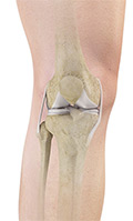
The knee is a complex joint made up of various structures including bones, tendons, ligaments and muscles. They all work together to maintain normal function and provide stability to the knee during movement.
Meniscus Tear
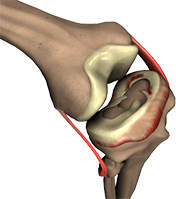
Meniscal tears are the most common knee injury in athletes, especially those involved in contact sports. A sudden bend or twist in your knee can cause the meniscus to tear. This is a traumatic meniscus tear. Older individuals are more prone to degenerative meniscal tears as the cartilage wears out and weakens with age. The two crescent shaped cartilage structures present between the femur and the tibia are called menisci. They stabilize the knee joint and act as "shock absorbers".
Patellofemoral Instability
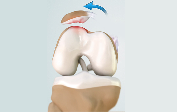
The knee can be divided into three compartments: patellofemoral, medial and lateral compartment. The patellofemoral compartment is the compartment in the front of the knee between the knee cap and thigh bone. The medial compartment is the area on the inside portion of the knee, and the lateral compartment is the area on the outside portion of the knee joint. Patellofemoral instability means that the patella (kneecap) moves out of its normal pattern of alignment.
Arthroscopy of The Knee Joint
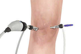
Knee Arthroscopy is a common surgical procedure performed using an arthroscope, a viewing instrument, to examine the knee joint and to diagnose or treat a knee problem. It is a very safe procedure and the patients are discharged home from the hospital on the same day of surgery.
Arthroscopic Meniscectomy
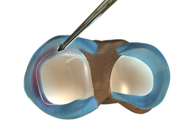
The arthroscope is a small fiber-optic viewing instrument made up of a tiny lens, light source and video camera. The surgical instruments used in arthroscopic surgery are very small (only 3 or 4 mm in diameter), but appear much larger when viewed through an arthroscope.
Anterior Cruciate Ligament ACL Reconstruction
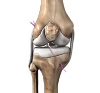
The anterior cruciate ligament is one of the major stabilizing ligaments in the knee. It is a strong rope like structure located in the center of the knee running from the femur to the tibia. When this ligament tears, it does not usually heal and often leads to the feeling of instability in the knee.